
The Genetics Society of AustralAsia encompasses research ranging from the micro to the macro scale. The following are some images of the science our members are conducting.
")
Drosophila ovary (Image © Anabel Herr)
")
DNA sequence alignments (Image © Anna MacDonald)
")
Chromosome spread from a cancer cell line (Image © Owen Marshall)
")
Fruit bodies of the holotype of Entoloma ravinensis in situ (Image © David Catcheside)

Aedes aegypti mosquito marked with fluorescent powder
© Perran Stott-Ross
")
DNA sequencing traces (Image © Anna MacDonald)

Drosophila alimentary canal expressing stress pathways in response to insecticides.
© Felipe Martelli Soares da Silva

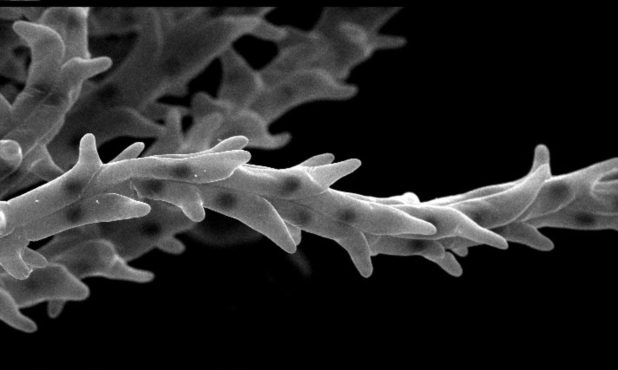
Electron microscopy of Drosophila retina exposed to insecticides
© Felipe Martelli Soares da Silva

Confocal micrograph of a Drosophila eye
© Felipe Martelli Soares da Silva

Thorax closure during metamorphosis in Drosophila
© Enoch Wong, 2018
")
Inside the egg – Development of Acritoscincus duperreyi (Eastern three-lined skink)
© Duminda Dissanayake
Nuclear Galaxy: Nucleus stained with DAPI and an
epigenetic modifier protein Smchd1, acquired using 3D structural illumination microscope.
© Iromi Wanigasuriya

Tornado: 10-day old wildtype adult Drosophila melanogaster midgut in the anterior region
© Fionna Zhu

3D image of an 8-month-old Tasmanian devil, reconstructed based on whole-body CT scans
©Yuanyuan Cheng / Australasian Wildlife Genomics Group

Cells of a wheat stigma
© Vy Nguyen

Penguins
© Tess Cole, 2018. Tess Cole's research focusses on the evolution of penguins. This image was inspired by several trips to Antarctica. Watercolour pencil and black ink.

Koscuiscola tristis male grasshopper thermoregulating.
© Sonu Yadav

Koscuiscola tristis resting on a bush
© Sonu Yadav
")
Koscuiscola tristis mating pair (female larger, male smaller)
© Sonu Yadav
")
Bone and cartilage in a 40-day old zebrafish (Danio rerio)
© Melanie Stewart, 2018

An optical section of a Drosophila melanogaster larval brain showing glia-neuron interactions
© Marta Portela

Drosophila melanogaster eye imaginal disc
© Marta Portela

Salivary glands from a Drosophila melanogaster larva
© Marta Portela

An optical section of a Drosophila melanogaster larval brain where a glioblastoma has been induced to study the cellular and molecular characteristics of the tumour
© Marta Portela

Image of Telmatactis autraliensis under blue light showing expression of GFP-like fluorescent chromoprotein in tentacles.
© Lauren Ashwood
")
A heart in the wrong place (ectopic Adb-B expression in the wing disc)
© Isabelle Lohrey, The University of Melbourne, 2019
 basking in the wild.")
A spotted skink (Carinascincus ocellatus) basking in the wild.
© Charles Foster, 2018

Functional resurrection of DNA under convergent positive selection in the Tasmanian tiger and grey wolf
© Laura E Cook, 2019