
The following images were submitted by GSA students and postdocs / early career researchers (ECRs) for the 2019 GSA Image Competition. The winner and runner up will be announced at the end of the 2019 GSA Meeting.

Functional resurrection of DNA under convergent positive selection in the Tasmanian tiger and grey wolf
© Laura E Cook, 2019

Slimb is required for cell pigmentation and cell survival
© Bichao Zhang, 2019
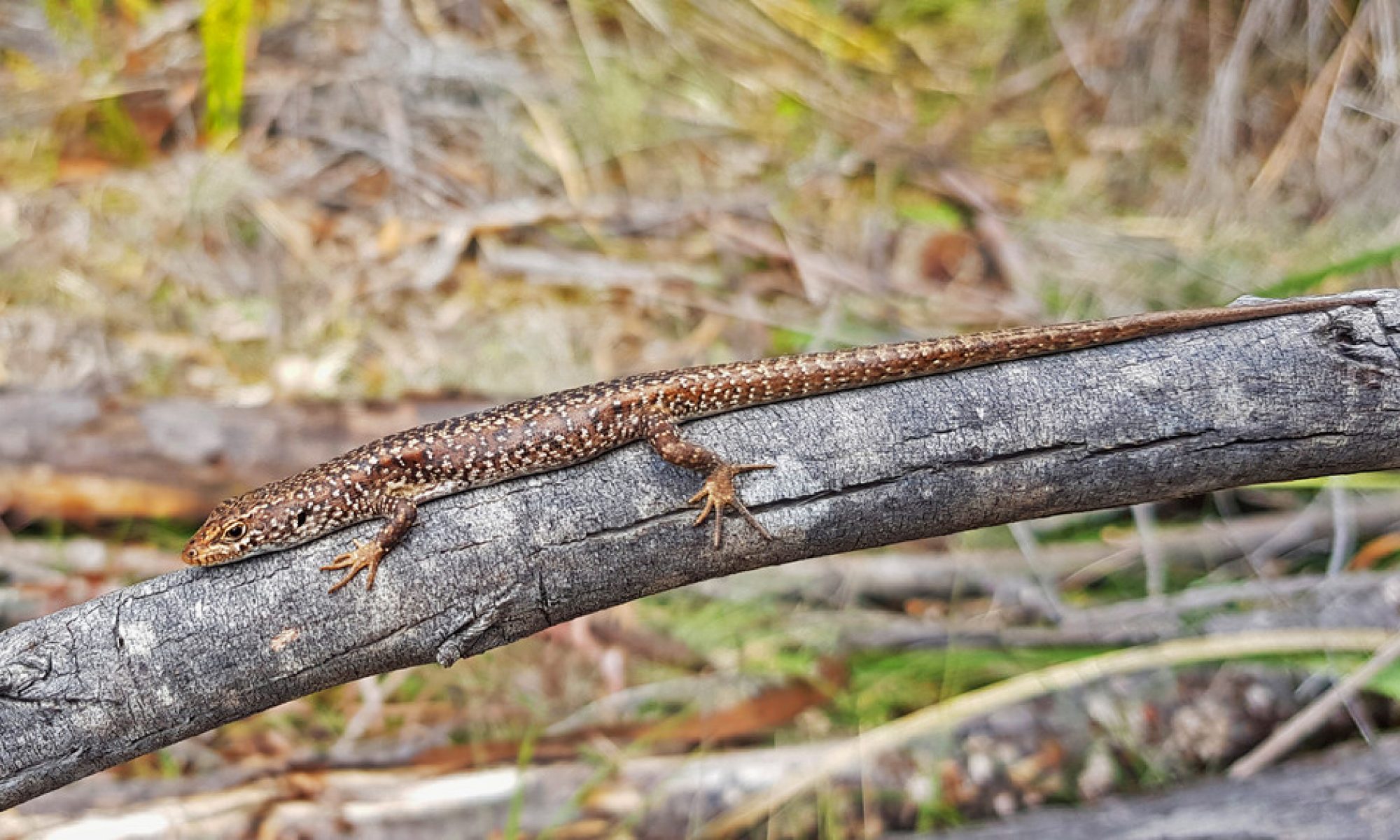
 basking in the wild.")
A spotted skink (Carinascincus ocellatus) basking in the wild.
© Charles Foster, 2018
")
A heart in the wrong place (ectopic Adb-B expression in the wing disc)
© Isabelle Lohrey, The University of Melbourne, 2019

Image of Telmatactis autraliensis under blue light showing expression of GFP-like fluorescent chromoprotein in tentacles.
© Lauren Ashwood

An optical section of a Drosophila melanogaster larval brain where a glioblastoma has been induced to study the cellular and molecular characteristics of the tumour
© Marta Portela

Salivary glands from a Drosophila melanogaster larva
© Marta Portela

Drosophila melanogaster eye imaginal disc
© Marta Portela

An optical section of a Drosophila melanogaster larval brain showing glia-neuron interactions
© Marta Portela

Dividing testis cell
© Melanie Stewart, 2018
")
Bone and cartilage in a 40-day old zebrafish (Danio rerio)
© Melanie Stewart, 2018
")
Bone and cartilage in a 40-day old zebrafish (Danio rerio)
© Melanie Stewart, 2018

A photo of Australian Alps taken in Kosciuszko national park
© Sonu Yadav
")
Koscuiscola tristis mating pair (female larger, male smaller)
© Sonu Yadav

Koscuiscola tristis resting on a bush
© Sonu Yadav

Koscuiscola tristis male grasshopper thermoregulating.
© Sonu Yadav

Penguins
© Tess Cole, 2018. Tess Cole's research focusses on the evolution of penguins. This image was inspired by several trips to Antarctica. Watercolour pencil and black ink.

The development of Arabidopsis thaliana ovule from two nucleate stage up to early globular stage
© Vy Nguyen

Inside a wheat pollen grain
© Vy Nguyen

Cells of a wheat stigma
© Vy Nguyen

Wheat stigma cells and pollens
© Vy Nguyen

3D image of an 8-month-old Tasmanian devil, reconstructed based on whole-body CT scans
©Yuanyuan Cheng / Australasian Wildlife Genomics Group

Confocal micrograph of a Drosophila eye
© Felipe Martelli Soares da Silva

Electron microscopy of Drosophila retina exposed to insecticides
© Felipe Martelli Soares da Silva

Drosophila alimentary canal expressing stress pathways in response to insecticides.
© Felipe Martelli Soares da Silva

Aedes aegypti mosquito marked with fluorescent powder
© Perran Stott-Ross

An Aedes aegypti mosquito marked with blue dye
© Perran Stott-Ross

Tornado: 10-day old wildtype adult Drosophila melanogaster midgut in the anterior region
© Fionna Zhu
Nuclear Galaxy: Nucleus stained with DAPI and an
epigenetic modifier protein Smchd1, acquired using 3D structural illumination microscope.
© Iromi Wanigasuriya
")
Inside the egg – Development of Acritoscincus duperreyi (Eastern three-lined skink)
© Duminda Dissanayake

Thorax closure during metamorphosis in Drosophila
© Enoch Wong, 2018